

Files
Download Full Text (1.8 MB)
Presentation date
Summer 8-7-2025
College, Institute, or Department
Anesthesiology
Faculty Mentor
Hanjun Wang
Research Mentor
Hanjun Wang
Abstract
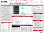
Background Genetic mutations that create pyruvate dehydrogenase complex (PDHc) deficiencies are a primary cause of congenital lactic acidosis (CLA). CLA is a rare genetic disorder in which the mitochondrion is unable to effectively metabolize excess amounts of pyruvate and lactate. It is estimated that ~250 – 300 live births per 1,000 are affected in the U.S each year. Mutations found in the X-linked gene for the E1α subunit of pyruvate dehydrogenase (pdha1) account for the majority of PDHc deficiency conditions. As pyruvate dehydrogenase acts to convert pyruvate into Acetyl CoA to be further utilized in generating ATP within the mitochondria, disruptions in this process can result in an ATP energy crisis, which acts as a contributing factor in the pathogenesis of a range of disease conditions.
Objectives A transgenic model was created to study the effects of the loss of the E1 subunit of pyruvate dehydrogenase and its subsequent effects on cardiac remodeling and cardiac glucose metabolism.
Methods Adult female Pdha1flox8 mice were bred to adult male αMHC-MerCreMer mice to create a tamoxifen-inducible knock-out of the pdha1 gene within cardiomyocytes. Cre-recombinase directed deletion of pdha1 was carried out by intraperitoneal injections (0.1mL/mouse) of Tamoxifen or Vehicle (15% ethanol in sunflower oil) in αMHC-MerCreMer:Pdha1flox/flox mutant mice at 16 weeks of age. Transthoracic echocardiography was performed on anesthetized mice at baseline (prior to injection), and the 1-month, 2-month, and 3-month timepoints following IP injection. Mitochondrial respiration measurements were obtained in 0.5 – 2 mg of permeabilized cardiac muscle fibers. A substrate-uncoupler-inhibitor titration (SUIT) protocol was performed to examine mitochondrial oxygen consumption at different respiratory states Cardiac tissues were collected from the pdha1 KO and vehicle mice at 3 months to perform western blot analysis to validate various metabolic and mitochondrial protein expression.
Results Tamoxifen induction of the pdha1 knock-out within cardiomyocytes revealed a time course of progressive cardiac dysfunction tracked by structural and functional changes of the heart through echocardiography. Parameters of both systolic and diastolic function are severely impacted at 3 months post tamoxifen, as shown by an average ejection fraction of 18.4%. Measurements of mitochondrial oxygen consumption revealed a decline in oxidative phosphorylation capacity and CI-linked respiration at 1 month post tamoxifen that preceded significant changes in hypertrophic remodeling and diminished cardiac function that ultimately resulted in heart failure. Initial western blot analysis confirmed the validity of the transgenic model used, but no other protein expression differed significantly between vehicle and pdha1 KO animals, aside from significantly decreased expression of SERCA2a, an ATP-dependent Ca2+ pump responsible for muscle relaxation; this was likely impacted from a loss of contractility and greater stiffness in the failing cardiac muscle.
Keywords
Pyruvate Dehydrogenase, Heart Failure, Mitochondria, Congenital Lactic Acidosis
Recommended Citation
Schulte, Collin T.; Nielson, Grace K.; Wilcox, Reesa M.; and Wang, Han-Jun, "Cardiac-Specific Knock-out of PDHA1 Leading to Mitochondrial Dysfunction and Progressive Heart Damage" (2025). Posters: 2025 Summer Undergraduate Research Program. 2.
https://digitalcommons.unmc.edu/surp2025/2